

 I'm A Patient
 I'm A Provider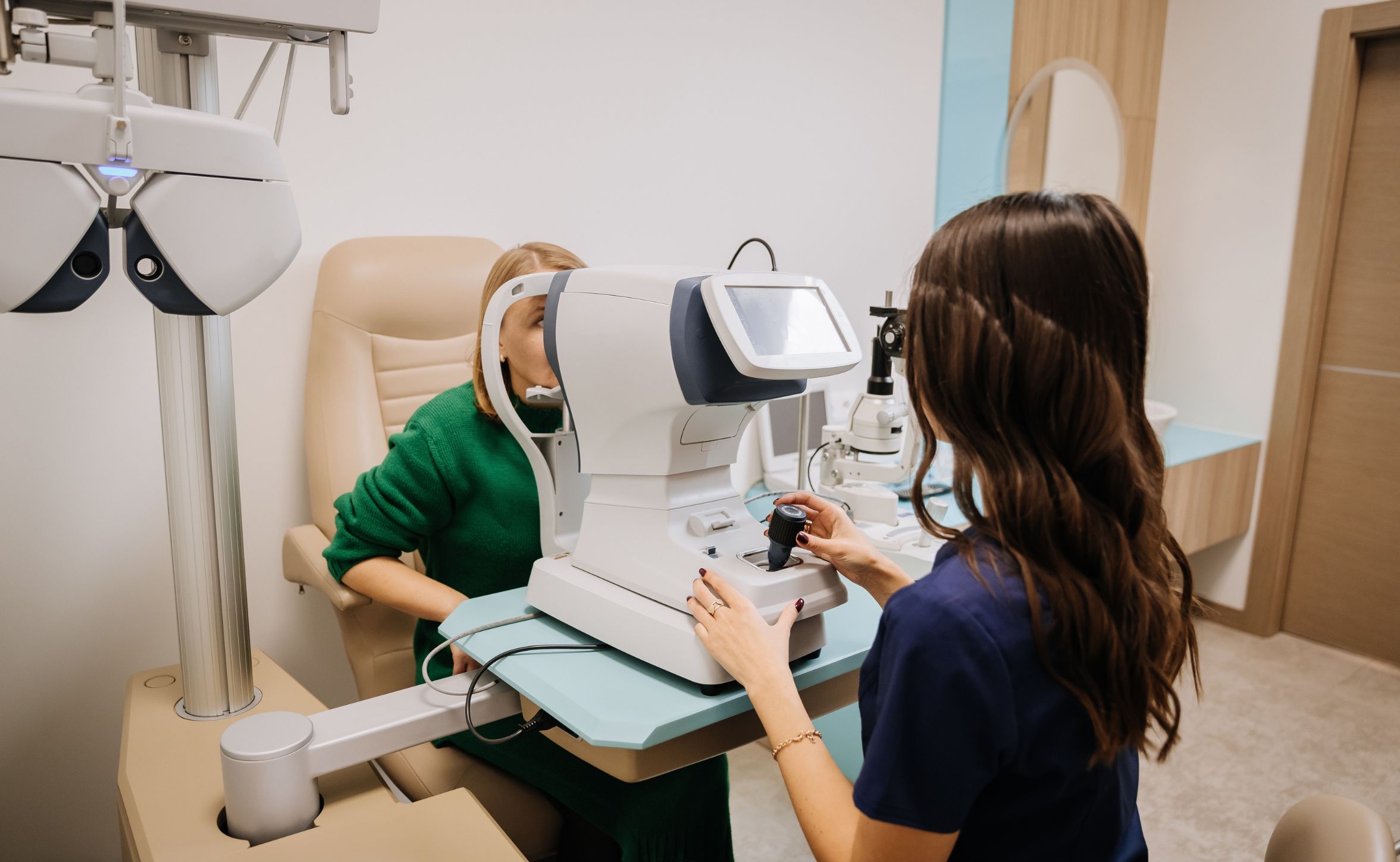
Direct Answer
Optometry clinics offering comprehensive dry age-related macular degeneration (AMD) care need equipment for both diagnosis and management. The diagnostic side requires optical coherence tomography (OCT) for retinal imaging, Early Treatment Diabetic Retinopathy Study (ETDRS) visual acuity charts for standardized acuity measurement, contrast sensitivity testing capability, and fundus photography for documentation. The treatment side, for clinics that offer device-based therapy, requires a Health Canada–authorized treatment device with a current regulatory licence for dry AMD.1
Each piece of equipment serves a distinct purpose. OCT provides the structural picture of the macula. ETDRS acuity and contrast sensitivity testing provide the functional picture. Fundus photography documents the clinical record over time. A Health Canada–authorized treatment device extends the clinic’s role from monitoring and advising to actively improving visual function in eligible patients.2
Why Equipment matters in Dry AMD Care
Dry AMD is a long-term condition that requires consistent, well-documented monitoring over years. The quality of clinical decisions at each stage depends directly on the quality of the information available. A clinic that can only assess visual acuity with a standard Snellen chart and has no retinal imaging capability is not in a position to accurately stage dry AMD, reliably detect structural changes, or make informed decisions about treatment eligibility.3
As the range of management options for dry AMD has expanded, the equipment requirements for a complete dry AMD service have also grown. A clinic that can monitor dry AMD but cannot offer or refer for device-based therapy is offering an incomplete service to patients who may be candidates for functional improvement. Conversely, a clinic that offers treatment without the diagnostic infrastructure to confirm eligibility and track outcomes is not using the treatment appropriately.1
The sections below cover each equipment category in terms of what it measures, why it matters for dry AMD care and what to look for when evaluating options.
Diagnostic Equipment
Optical Coherence Tomography (OCT)
OCT is the single most important piece of imaging equipment for an AMD clinic. It produces high-resolution cross-sectional images of the macula that allow the clinician to visualize the layers of the retina, quantify drusen volume and distribution, identify areas of retinal pigment epithelium atrophy, and detect signs of subretinal fluid or neovascularisation that may indicate conversion from dry to wet AMD.1
Without OCT, AMD staging relies on clinical examination and fundus photography alone, which do not provide the structural detail needed to accurately track disease over time or to detect early conversion to wet AMD. For any practice managing more than a small number of AMD patients, OCT is essential, not optional.1
Spectral domain OCT is the current standard for retinal imaging in clinical practice. Swept-source OCT offers deeper penetration and wider-field imaging and is increasingly available at practice level. The choice of system should be guided by image quality, ease of use, software integration with patient records, and the availability of normative databases and progression analysis tools.
OCT-A, or OCT angiography, provides additional information about retinal vasculature without the need for dye injection and is particularly useful for detecting early choroidal neovascularisation. It is a valuable supplementary tool for practices managing patients at risk of wet AMD conversion, though it does not replace standard OCT for routine monitoring.1
ETDRS Visual Acuity Charts
The Early Treatment Diabetic Retinopathy Study chart is the standard for visual acuity measurement in AMD clinical trials and is the appropriate tool for AMD monitoring in a clinical setting. Unlike the traditional Snellen chart, which groups letters in rows of decreasing size, the ETDRS chart presents letters in a standardized format with equal numbers per row and consistent spacing, allowing letter-by-letter scoring that is sensitive to small changes over time.2
This sensitivity matters in AMD care because the clinical question is often not whether vision has changed dramatically but whether it has changed at all. A standardized ETDRS protocol, conducted at a fixed test distance with consistent lighting and appropriate refraction, can provide a reliable baseline against which future measurements can be compared. A standard Snellen chart does not offer the same precision.
Clinics using authorized device-based therapy for AMD should use ETDRS acuity measurement as the standard outcome tracking tool, consistent with the methodology used in the device’s clinical trial. This allows meaningful comparison of individual patient outcomes against published trial results.
Contrast Sensitivity Testing
Contrast sensitivity, the ability to detect objects against backgrounds of varying contrast, is frequently reduced in dry AMD independently of ETDRS visual acuity. A patient who retains relatively good high-contrast acuity may still experience significant difficulty with real-world visual tasks such as reading in low light, identifying faces in variable lighting or navigating environments where contrast is reduced. Contrast sensitivity testing captures this dimension of visual function that acuity alone misses.3
In the context of device-based therapy, contrast sensitivity is a co-primary outcome measure alongside ETDRS acuity in published trials. MacuMira’s clinical trial measured contrast sensitivity in log units and reported an average improvement of 0.24 log units in the treatment group. A clinic that tracks treatment outcomes without measuring contrast sensitivity is missing one of the two primary endpoints used to evaluate treatment effectiveness.4
Several validated contrast sensitivity testing systems are available for clinical use. The Pelli-Robson chart, Mars Letter Contrast Sensitivity test and computerized systems such as the CSV-1000 are commonly used in AMD research and clinical practice. Whichever system is adopted, consistent methodology, standardized test conditions, and calibrated equipment are important for producing reliable longitudinal data.
Fundus Photography
Fundus photography provides a two-dimensional colour image of the posterior pole and is used for documenting drusen distribution, retinal pigment epithelium changes, and geographic atrophy. While it provides less structural detail than OCT, it serves an important documentation function and allows straightforward visual comparison of the posterior pole over time.1
Wide-field fundus photography extends the field of view beyond the central macula and can detect peripheral retinal changes that may be relevant to differential diagnosis or disease monitoring. Ultra-wide-field systems covering 200 degrees or more are increasingly available at the practice level, though for AMD monitoring specifically, the central macula is the area of primary clinical concern.
Fundus photography is also useful for patient communication. Showing a patient a colour image of their macula and how it has changed over time is often more accessible than explaining an OCT cross-section and can support better patient engagement with their monitoring and management plan.
Fundus Autofluorescence
Fundus autofluorescence (FAF) imaging uses the natural fluorescence of lipofuscin in the retinal pigment epithelium to produce images that highlight areas of RPE health and dysfunction. In dry AMD, FAF is particularly useful for mapping the extent and boundaries of geographic atrophy, which appears as a dark area of absent autofluorescence surrounded by a hyperautofluorescent border in active disease.1
FAF is not universally available in community optometry but is increasingly integrated into multimodal imaging platforms alongside OCT and colour fundus photography. For practices managing patients with advanced dry AMD or geographic atrophy, access to FAF imaging is a meaningful addition to the diagnostic toolkit.
Treatment Equipment
Authorized Device-Based Therapy for Dry AMD
For clinics that want to offer active treatment rather than limit their AMD services to monitoring and supplementation, a Health Canada–authorized treatment device to improve visual function is an essential addition. In Canada, the device must hold a Health Canada Medical Device Licence for dry AMD. In Australia, TGA registration. Any device offered for clinical use should also be backed by a published randomized controlled trial with a sham comparator and validated outcome measures.2
MacuMira is a Health Canada–authorized treatment device indicated to improve visual function in patients with dry AMD. It delivers low-level electrical stimulation noninvasively to tissues near the eye in a clinical setting under the supervision of a licensed eye care professional. In its published randomized sham-controlled trial, the treatment group showed statistically significant improvement in both visual acuity and contrast sensitivity. The average gain was 8.2 letters on the ETDRS scale, with 48% of treated patients gaining 10 or more letters. Contrast sensitivity improved by an average of 0.24 log units. Zero device-related serious adverse events were recorded in the trial or across more than 21,000 real-world treatments in 3 countries.4
MacuMira is not indicated for wet AMD. Treatment eligibility requires a clinical assessment including confirmation of dry AMD stage and baseline visual acuity. The published trial enrolled patients with visual acuity between 20/50 and 20/200. Patients with better baseline acuity may experience a ceiling effect that limits the measurable scope for improvement.4
From an equipment and workflow perspective, integrating a treatment device into an AMD clinic requires planning for appointment scheduling, staff training in the treatment protocol and a systematic approach to outcome documentation. Clinics should establish a standard protocol for measuring and recording ETDRS acuity and contrast sensitivity before each treatment course and at defined intervals thereafter.
Building a Complete AMD Service: Equipment and Workflow Together
The clinical value of each piece of equipment is greatest when it functions as part of an integrated workflow rather than in isolation. OCT findings inform treatment eligibility decisions. ETDRS and contrast sensitivity baselines set the reference point against which treatment outcomes are measured. Fundus photography provides the longitudinal visual record. The treatment device delivers the intervention. Together they create a service that can assess, treat, and track AMD patients within a single practice.1
Practices building out an AMD service should also consider the referral pathways that connect their capabilities to those of other providers. Patients who convert from dry to wet AMD need prompt access to anti-VEGF treatment through an ophthalmologist or retinal specialist. Patients with advanced geographic atrophy may benefit from low vision rehabilitation referral. Having clear and established pathways for these situations is part of providing comprehensive AMD care, not only having the right equipment.
Questions Optometrists Should Ask when Equipping an AMD Clinic
- Does the OCT system I am considering provide the resolution, software tools and progression analysis needed for longitudinal AMD monitoring?1
- Is my visual acuity testing protocol ETDRS-standardized or do I need to update my equipment and methodology?
- What contrast sensitivity testing system is most appropriate for my patient volume and the outcome tracking I need to do?3
- Does the treatment device I am considering hold a current regulatory licence for dry AMD in the countries where I practice?2
- Have I read the primary clinical trial publication for the device and do the eligibility criteria match my patient population?4
- Do I have a clear protocol for documenting baseline and follow-up outcomes for patients receiving device-based therapy?
- Are my referral pathways for wet AMD conversion and low vision rehabilitation clearly established?
Key Takeaway
A comprehensive optometry AMD clinic needs OCT for retinal imaging, ETDRS acuity charts for standardized visual acuity measurement, contrast sensitivity testing for functional assessment, fundus photography for longitudinal documentation, and an authorized treatment device for clinics offering visual function improvement therapy.
MacuMira is a Health Canada–authorized treatment device indicated to improve visual function in dry AMD patients, supported by a published randomized sham-controlled trial and real-world use across more than 21,000 treatments with zero device-related serious adverse events. It is for use under the supervision of a licensed eye care professional and is not indicated for wet AMD.
The most effective AMD clinic integrates all of these capabilities into a coordinated workflow with clear referral pathways for situations that fall outside the primary care optometry scope.
Disclaimer
This article is for informational and educational purposes only. Equipment recommendations are general in nature and do not constitute endorsement of any specific manufacturer or product. Clinicians should apply their own professional judgment when selecting equipment for their practice. Patients should consult a qualified eye care professional for diagnosis and treatment of dry AMD.
Glossary
Optical coherence tomography (OCT): A noninvasive retinal imaging technology producing high-resolution cross-sectional images of the macula. The standard tool for AMD staging and progression monitoring.
OCT angiography (OCT-A): An extension of OCT technology that images retinal vasculature without dye injection. Useful for detecting early choroidal neovascularisation associated with wet AMD conversion.
ETDRS scale: The Early Treatment Diabetic Retinopathy Study visual acuity scale, scored letter by letter at a standardized test distance. More sensitive to small changes than Snellen notation.
Contrast sensitivity: The ability to distinguish objects from their background at varying contrast levels. Frequently reduced in dry AMD independently of high-contrast visual acuity.
Fundus autofluorescence (FAF): An imaging modality that uses natural fluorescence of lipofuscin in the retinal pigment epithelium to highlight areas of RPE health and dysfunction, including the extent of geographic atrophy.
MacuMira: A Health Canada–authorized treatment device indicated to improve visual function in patients with dry AMD, delivered via low-level electrical stimulation in a clinical setting under the supervision of a licensed eye care professional. Not indicated for wet AMD.
Geographic atrophy: An advanced stage of dry AMD in which large areas of retinal pigment epithelium and photoreceptors have degenerated, causing irreversible central vision loss.
Ceiling effect: In clinical measurement, the limited scope for measurable improvement when a patient’s baseline function is near the upper end of the measurement scale.
Dry AMD: A progressive retinal condition affecting the macula. The most common form of AMD. Distinct from wet AMD in pathophysiology and management.
Wet AMD: A form of AMD involving abnormal blood vessel growth beneath the retina. Treated with anti-VEGF injections. MacuMira is not indicated for wet AMD.
References
1. Age-Related Macular Degeneration. National Eye Institute, National Institutes of Health. Last reviewed November 2023. Accessed June 2026. https://www.nei.nih.gov/eye-health-information/eye-conditions-and-diseases/age-related-macular-degeneration.
2. Medical Device Licences. Health Canada. Date not listed. Accessed June 2026. https://www.canada.ca/en/health-canada/services/drugs-health-products/medical-devices/licences.html.
3. Understanding Macular Degeneration. American Academy of Ophthalmology. Published November 2025. Accessed June 2026. https://www.aao.org/eye-health/diseases/amd-macular-degeneration.
4. Evaluation of visual acuity in dry AMD patients after microcurrent electrical stimulation. Parkinson KM, Sayre EC, Tobe SW. International Journal of Retina and Vitreous. Published May 2023. Accessed June 2026. https://macumira.com/wp-content/uploads/2025/07/MacuMira-Clinical-Trial.pdf.
5. Age-Related Macular Degeneration (AMD). Canadian Ophthalmological Society. Date not listed. Accessed June 2026. https://www.seethepossibilities.ca/eye-health/age-related-macular-degeneration/.